A comprehensive assessment demands both knowledge and time, distinguishing objective signs from subjective findings like pain and tenderness․
Understanding anatomy is crucial, alongside observing posture, gait, and movement during the physical exam, especially in older adults․
Importance of a Thorough Examination
A meticulous musculoskeletal examination is paramount for accurate diagnosis and effective treatment planning․ Distinguishing between objective findings – swelling, limited range of motion – and subjective reports of pain is vital․
Systemic disorders can manifest as musculoskeletal complaints, necessitating a broad assessment․ Identifying potential “red flags,” like pain at rest, prompts further investigation․ A complete evaluation considers historical clues, onset, location, and functional limitations to guide clinical decision-making․
Anatomical Knowledge as a Foundation
Performing an accurate and meaningful musculoskeletal examination fundamentally relies on a robust understanding of anatomy․ Having an atlas readily available – either physical or digital – serves as a quick reference during the assessment process․
This foundational knowledge enables precise palpation, range of motion assessment, and interpretation of findings, ultimately leading to a more informed and effective clinical evaluation of the patient’s condition․

Patient History: Gathering Crucial Clues
Historical clues regarding onset, location, radiation, severity, and modifying factors are vital, alongside functional limitations and prior problems․
Onset, Location, and Radiation of Pain
Determining the pain’s onset – whether acute or slowly progressive – is fundamental․ Precisely pinpointing the location and understanding if the pain radiates elsewhere provides critical diagnostic clues․
Was there a specific injury mechanism? Exploring these details helps differentiate between traumatic and non-traumatic causes․ A thorough inquiry into these aspects forms the basis for focused examination and accurate diagnosis, guiding further assessment․
Severity and Modifying Factors
Assessing pain severity is crucial, but equally important is identifying what makes it better or worse․ Understanding these modifying factors – activities, positions, or treatments – offers valuable insights․
Detailed questioning about symptom relief or exacerbation helps refine the differential diagnosis․ This information directs the physical examination, focusing on provocative maneuvers and assessing functional limitations effectively․
Functional Limitations and Impact
Determining the extent to which musculoskeletal complaints affect daily activities is paramount․ Explore specific limitations – walking, lifting, dressing – and how symptoms impact the patient’s quality of life․
Understanding these functional consequences guides rehabilitation planning and establishes realistic goals․ Assess whether symptoms affect a single joint or multiple areas, and if the onset was acute or gradual․

Observation: Initial Assessment
Begin by carefully observing the patient’s posture, gait, and extremity movements during the exam, noting any visible asymmetries or deformities․
Posture and Gait Analysis
Assessing posture involves observing the patient from multiple angles – anterior, posterior, and lateral – to identify any deviations from normal alignment․ Note any spinal curvatures, shoulder height discrepancies, or pelvic tilt․
Gait analysis requires observing the patient’s walking pattern, paying attention to stride length, arm swing, and any limping or unusual movements․ Observe for smoothness, symmetry, and any signs of pain or instability during ambulation․
Symmetry and Deformities
Careful observation for symmetry is paramount; compare both sides of the body for any noticeable differences in limb length, muscle bulk, or joint alignment․ Look for visible deformities such as swelling, angulation, or dislocations․
Palpate bony landmarks to confirm visual findings and identify any subtle asymmetries․ Document any observed deformities precisely, noting their location, size, and any associated tenderness or functional limitations․
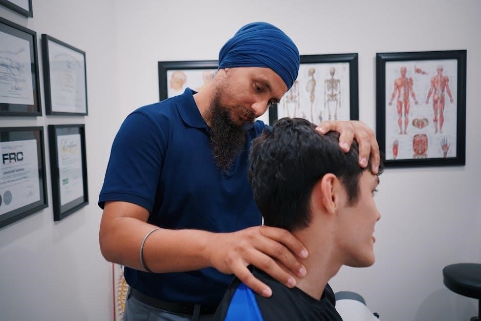
Palpation: Detecting Abnormalities
Palpate muscles for tenderness and spasm, and joints for swelling or warmth, carefully distinguishing objective versus subjective findings during assessment․
Muscle Palpation for Tenderness and Spasm
Systematic muscle palpation is essential, assessing for localized tenderness, involuntary muscle spasm, or palpable masses․ Utilize a gentle, circular motion, comparing bilaterally to identify asymmetries․ Note any areas eliciting pain or resistance, documenting the location and intensity precisely․
Consider muscle bulk and tone; atrophy or hypertrophy can indicate chronic disuse or overuse․ Palpation helps differentiate between muscular and joint origins of pain, guiding further examination․
Joint Palpation for Swelling and Warmth
Carefully palpate joints for effusion, swelling, and increased warmth, comparing to the contralateral side․ Assess for crepitus during gentle range of motion․ Note any localized tenderness over joint lines or bony prominences, indicating potential inflammation or injury․
Distinguish between intra-articular and extra-articular findings; swelling suggests effusion, while warmth can indicate inflammation․ Palpation aids in identifying joint instability or structural abnormalities․
Range of Motion (ROM) Assessment
Assess both active and passive ROM, meticulously documenting measurements to identify limitations or pain․ This reveals functional impairments and guides treatment․
Active vs․ Passive ROM
Active range of motion involves the patient moving a joint independently, demonstrating muscle strength and neurological function․ Passive ROM is when the examiner moves the joint, assessing the joint’s structural integrity and any limitations not caused by muscle weakness․
Comparing these two reveals crucial information; pain during active motion suggests muscle or tendon issues, while pain during passive motion points to joint problems․ Documenting both accurately is essential for a complete musculoskeletal evaluation․
Documenting ROM Measurements
Accurate documentation of range of motion (ROM) is vital, typically recorded in degrees using a goniometer․ Note the type of movement (flexion, extension, abduction, etc․) and compare to normative values or the unaffected side․
Clearly record any limitations, pain points, or muscle guarding observed during the assessment․ Consistent and precise documentation facilitates tracking progress and informs treatment plans, ensuring comprehensive patient care and effective communication․

Muscle Strength Testing
Assess strength using a standardized grading system, evaluating common muscle groups to identify weaknesses or imbalances impacting function and mobility․
Grading Muscle Strength
Muscle strength is typically graded on a scale from 0 to 5, providing a standardized method for documenting functional ability․ A grade of 0 indicates no contraction, while 5 represents normal strength against gravity and resistance․
Intermediate grades denote varying degrees of contraction; 1 is a trace contraction, 2 is movement with gravity eliminated, 3 is movement against gravity, and 4 is movement against gravity with some resistance․ Accurate grading requires stabilization and consistent application of resistance․
Common Muscle Groups to Assess
During a musculoskeletal examination, assessing key muscle groups is essential for identifying weakness or imbalances․ Common groups include shoulder abduction and adduction, elbow flexion and extension, wrist flexion and extension, hip flexion, knee extension and flexion, and ankle dorsiflexion and plantarflexion․
Testing these groups provides valuable insight into nerve function and muscle integrity, helping pinpoint potential areas of concern and guiding further diagnostic evaluation․

Special Tests for Specific Conditions
Specific tests help diagnose ligamentous injuries and meniscal tears, complementing the broader musculoskeletal assessment for accurate diagnoses․
Tests for Ligamentous Injuries
Ligamentous stability is assessed through specific provocative maneuvers, stressing the joint to identify laxity or pain indicating damage․ These tests evaluate the integrity of crucial ligaments, such as those found in the knee – anterior cruciate (ACL), posterior cruciate (PCL), medial collateral (MCL), and lateral collateral (LCL) – and shoulder․
Examples include the Lachman test and anterior drawer test for ACL integrity, and varus/valgus stress tests for collateral ligament stability․ Careful observation and palpation during these tests are vital for accurate interpretation․
Tests for Meniscal Tears
Evaluating for meniscal tears involves tests designed to elicit pain or mechanical symptoms, like clicking or locking, within the joint․ The McMurray test is a classic maneuver, involving joint flexion and rotation while palpating for a palpable snap or pain along the joint line․
Apley’s compression and distraction tests also help differentiate meniscal from ligamentous injuries․ Positive findings suggest a potential tear, warranting further imaging like MRI for confirmation and precise localization․

Neurological Examination of Musculoskeletal System
Assessing reflexes and sensory function is vital, as nerve compression or injury can mimic musculoskeletal issues, impacting motor control and sensation․
Assessment of Reflexes
Reflexes provide crucial insights into the integrity of the nervous system’s pathways influencing musculoskeletal function․ Testing typically involves deep tendon reflexes – biceps, triceps, brachioradialis, patellar, and Achilles – using a reflex hammer․
Grading reflexes on a scale (0-4+) helps identify abnormalities; diminished or absent reflexes suggest nerve damage, while hyperreflexia can indicate upper motor neuron lesions․ Observing symmetry is key, as asymmetry warrants further investigation․
Sensory Examination
A thorough sensory exam assesses the patient’s ability to perceive light touch, pain, temperature, and proprioception within the dermatomal distribution of relevant nerves․ This helps identify potential nerve compression or damage affecting musculoskeletal function․
Testing involves lightly touching the skin, using a sharp/dull object, and assessing joint position sense․ Documenting any sensory deficits – numbness, tingling, or altered sensation – is crucial for accurate diagnosis and treatment planning․
Considerations for Older Adults
Be mindful of age-related degeneration, limited mobility, and muscle weakness; avoid causing pain or discomfort during the musculoskeletal examination process․
Age-Related Changes in Mobility
As individuals age, expect to encounter decreased joint flexibility and range of motion due to cartilage wear and potential osteoarthritis development․ Muscle mass naturally declines, contributing to weakness and impacting stability․ These changes can affect gait, balance, and overall functional capacity․
Therefore, a careful and gentle approach is essential during the musculoskeletal examination of older adults, acknowledging these inherent physiological alterations and potential limitations․
Avoiding Pain and Discomfort During Examination
When assessing older adults, prioritize their comfort and avoid exacerbating existing pain or limitations․ Gentle movements and careful palpation are crucial, respecting their reduced mobility and potential joint degeneration․
Never force range of motion or push through resistance, as this can cause unnecessary discomfort․ Communicate clearly, explaining each step and pausing if the patient experiences pain․

Distinguishing Objective vs․ Subjective Findings
Objective signs—swelling, limited ROM—differ from subjective ones like tenderness and pain; careful differentiation is vital for accurate musculoskeletal assessment․
Objective Signs: Swelling, Effusion, Limited ROM
Observable and measurable indicators constitute objective findings, providing concrete evidence of musculoskeletal issues․ Swelling, often visible and palpable, suggests inflammation or fluid accumulation within tissues․ Effusion, specifically joint swelling, can be detected through palpation or visual inspection․
Limited range of motion (ROM), objectively assessed during examination, indicates structural or functional impairments․ These signs, unlike subjective reports of pain, are independently verifiable by the examiner, bolstering diagnostic accuracy․
Subjective Signs: Tenderness, Pain
Subjective data relies on the patient’s perception and description of their symptoms, forming a crucial part of the musculoskeletal assessment․ Tenderness, elicited during palpation, indicates localized discomfort upon touch, revealing areas of potential tissue damage or inflammation․
Pain, a primary complaint, is characterized by intensity, location, and quality, as reported by the patient․ These experiences, while vital, are not directly observable and require careful interpretation alongside objective findings․

Musculoskeletal Complaints and Systemic Disorders
Musculoskeletal symptoms can signal underlying systemic issues affecting joints, muscles, and tendons; pain at rest suggests inflammation or neoplasm․
Identifying Systemic Causes of Musculoskeletal Pain
Recognizing musculoskeletal complaints as potential indicators of broader systemic disorders is vital during examination․ Conditions like rheumatoid arthritis, lupus, or even infections can manifest primarily through joint and muscle pain․ Therefore, a thorough history and physical should actively explore systemic symptoms – fever, fatigue, weight loss, or skin rashes – alongside localized findings․ Pain present at rest frequently points towards acute inflammatory, neurological, or neoplastic processes, demanding further investigation beyond a purely musculoskeletal focus․
Pain at Rest: Red Flags
Pain occurring even at rest serves as a significant warning sign, deviating from typical musculoskeletal discomfort․ This symptom often suggests an underlying acute inflammatory process, a neurological issue, or potentially a neoplastic condition requiring immediate attention․ Clinicians must diligently investigate such cases, considering possibilities beyond simple muscle strains or sprains, and promptly pursue appropriate diagnostic testing to rule out serious pathology․
Documentation and Reporting
Accurate and concise record-keeping is paramount, ensuring effective communication of findings to other healthcare professionals for optimal patient care․
Accurate and Concise Record Keeping
Detailed documentation of both objective and subjective findings is essential for tracking patient progress and informing treatment plans․ Records should clearly articulate observed swelling, range of motion limitations, and any elicited tenderness․ Precise descriptions of pain characteristics – onset, location, radiation, and modifying factors – are crucial․ Avoid vague terminology; instead, utilize standardized measurements and descriptive language to ensure clarity and facilitate effective communication among the healthcare team, ultimately supporting continuity of care․
Communicating Findings Effectively
Clearly conveying examination results to patients and colleagues is paramount for collaborative care․ Explain findings in understandable terms, avoiding medical jargon whenever possible․ Highlight both objective signs – like limited range of motion – and subjective reports of pain․ A concise summary, emphasizing functional limitations and potential diagnoses, ensures everyone is aligned․ Effective communication fosters trust and shared decision-making, leading to optimal patient outcomes and a well-coordinated treatment strategy․